How Safe Are X-Rays, MRIs, Mammograms, and CT Scans? What You Need to Know About Radiation and Health Risks
Is there a limit to the number of scans you can have in a year? Should you tell your doctor if you've had a recent scan, even if it's a dental X-ray?

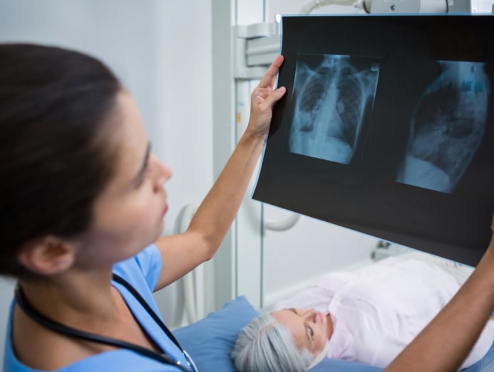
So you’re in the hospital. You’re worried about the medical emergency that brought you there. The doctor hasn’t said much, but has ordered some imaging tests like a chest x-ray and a CT scan.
Or you may be scheduled for a mammogram next week and just remember the results of a recent dental X-ray. In another case, you may be asked to have a PET scan after your annual checkup revealed something suspicious.
If you've been in one of these situations before, you may be wondering: Can you get too much radiation? Does it cause cancer? Do you need to speak up even if you're not pregnant?
HOW MUCH RADIATION ARE YOU EXPOSED TO?
“The amount of radiation used each time varies significantly,” said Associate Professor Lionel Cheng, senior consultant and head of the Department of Diagnostic Imaging at Singapore General Hospital.
It essentially depends on the type of imaging test being used. For example, the typical dose of radiation emitted during an X-ray, bone density scan or mammogram is lower than that emitted during a CT scan or PET scan, Associate Professor Cheng said.
According to Parkway Radiology, a simple dental, chest, or limb X-ray carries a negligible radiation risk of 1 in 1,000,000. Or the equivalent of several days of background radiation exposure. That’s right, you’re being exposed to natural and unavoidable radiation from the environment, like the ground, the air, building materials, and cosmic rays from outer space.
According to Parkway Radiology, even the so-called “higher” radiation from a CT scan or PET scan only carries a low cancer risk – 1 in 10,000 to 1 in 1,000, to be exact, or the equivalent of several years of exposure to natural radiation.
The area being photographed (for example, an arm versus the whole body) and the duration of the photo shoot also determine the amount of exposure, says Associate Professor Cheng.
IS THERE A LIMIT TO THE NUMBER OF SCANS YOU CAN DO IN A YEAR?
“There is no specific upper limit for the number of scans a patient can have,” said Associate Professor Cheng. “Some patients with complex or urgent conditions may need multiple scans in a short period of time. Other patients may only need one or two scans over several years.”
Rather than focusing on a specific number, “it's more important for patients to let their doctors know if they've had any other scans recently,” he says.

If you have had an ultrasound scan at a GP or public hospital in the past, your doctor will be able to access that record from the public healthcare system – and reduce duplicate tests and schedule the right follow-up ultrasound at the right time, says Associate Professor Cheng.
However, “scans performed in the private sector or overseas will not be in the doctor’s clinical record, so it is important that the patient provides that information,” he continued. “The doctor can then take this into account when considering any other medical imaging tests.”
WHY DO DOCTORS SOMETIMES ORDER MULTIPLE TYPES OF IMAGING TESTS?
Sometimes “a single scan doesn’t provide enough information to make an accurate diagnosis,” said Betty Matthew, a senior radiologist at SATA CommHealth.
“The use of multiple imaging modalities allows for a more comprehensive assessment, ensuring accurate diagnosis, effective treatment planning, and careful monitoring of the patient's condition.”
For example, an X-ray can help doctors detect broken bones from an accident but can’t check for internal bleeding and organ damage, which CT scans and MRIs can, Matthew said. She cited the following additional situations where multiple imaging tests may be needed:
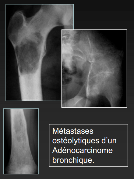
- Confirming the diagnosis: Take lung cancer, for example. A chest X-ray may show a lung mass, but a CT scan or MRI will provide a clearer picture. In stroke patients, a CT scan checks for bleeding in the brain, while an MRI assesses the extent of brain damage.
- Monitoring disease progression: Imaging tools such as PET, CT, and MRI are used to monitor the growth or spread of cancerous tumors. For patients with chronic diseases such as multiple sclerosis, repeated MRI scans may be needed to check for new lesions.
- Detecting infection or inflammation: Ultrasound, CT scan, or PET scan can help detect the source of infection.
SHOULD YOU WORRY ABOUT DENTAL X-RAYS?
Speak up if you’re pregnant. Otherwise, modern X-ray equipment limits the beam size to the area being scanned, the American Dental Association (ADA) says, so there’s no need for protection like a thyroid collar to protect against outside radiation exposure.
Following ADA guidelines, local dentists like Dr. Andrew Chia of Vista Dental Surgery also do not use thyroid collars because they “can accidentally block the X-ray beam, ruining the image,” he said. “X-rays need to be retaken.”
“When this happens, more X-rays are needed, and we want to avoid unnecessary X-rays,” said Professor Purnima Kumar, chair of the Department of Periodontics and Oral Medicine at the University of Michigan School of Dentistry and a member of the ADA Scientific Council.
HOW ARE THE DIFFERENT SCAN VERSIONS?
Why do you sometimes get a CT scan instead of an X-ray? Is the radiation from a mammogram higher than a regular X-ray? We find out:
1. X-RAY
What it is: Matthew says it uses a form of high-energy electromagnetic radiation to take detailed images of body structures. “Because X-rays involve ionizing radiation, exposure is carefully controlled and limited.”
Dr Lee Chau Hung, senior consultant at the Department of Diagnostic Imaging at Tan Tock Seng Hospital, added that X-rays have “very low radiation doses” and are “suitable for all ages and genders”.
When used: Matthew says it's commonly used to detect broken bones, dislocations, lung infections like pneumonia, and certain abdominal conditions.
Who’s not suitable: Pregnant women, as the fumes can harm the developing fetus. Otherwise, “there’s no absolute rule out X-rays,” Matthew says. However, X-rays are not prescribed indiscriminately and “are only recommended when the benefits outweigh the risks,” she says.
2. COMPUTED TOMATOGRAPHY (CT SCAN)
What it is: Think of it as a large, doughnut-shaped scanner. It emits multiple X-ray beams to create three-dimensional images of the inside of your body, Dr. Lee explains.
When to use it: “It’s very useful in providing detailed images of most internal organs.” And with modern technology, you can be scanned from head to toe in just one breath hold or less than 20 seconds, says Dr. Lee.
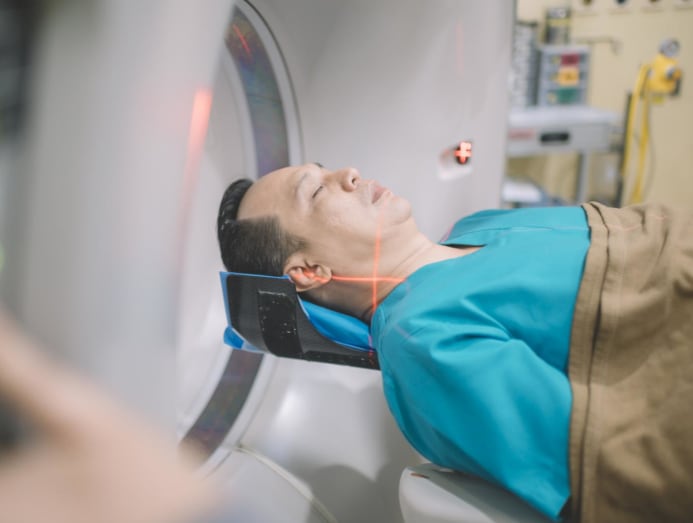
Who's not a good candidate: This method requires the highest dose of radiation and is generally not recommended for children, pregnant women, and teenagers unless absolutely necessary, says Dr. Lee.
Additionally, if you have asthma, allergies, or kidney disease, you may not be a good candidate for a CT scan. A dye or contrast agent is injected before the scan, and you may have a reaction to the dye, says Dr. Lee.
But it’s not a “no” because steroids can help reduce the risk of asthma attacks or allergic reactions. If not, they may be advised to have imaging tests instead, he said.
3. MAGNETIC RESONANCE IMAGING (MRI)
What it is: It’s also a large, ring-like scanner, but you’ll spend more time in it than a CT scanner. It generates electromagnetic waves to get “very detailed three-dimensional images of internal organs,” and has “the highest resolution of any imaging test,” according to Dr. Lee. But he also said it’s also one of the most expensive.
“Because it does not involve radiation, it is preferred over CT for children and adolescents. With modern technology, MRI is also safe during pregnancy,” said Dr. Lee.
Like a CT scan, dye or contrast material is injected to better visualize internal structures, says Dr. Lee. “Newer MRI contrast materials are very safe and can even be used in patients with kidney failure.”
When to use it: Dr. Lee says it’s typically used for very specific conditions. For example, doctors use it to look for nerve compression in the spine, small tumors in the liver and limbs, or small structures like the urinary tract and bile ducts.
Who it’s not for: Not for patients with claustrophobia or those who can’t hold still and monitor certain breathing patterns, Dr. Lee says. That’s because it takes a long time to get images. For example, an MRI of the lower back takes about 15 to 20 minutes, he says, while a liver scan can take up to 30 minutes.
“Most importantly, because MRI requires the use of high-intensity magnetic waves, it may not be safe for patients with metal implants such as clips, staples, cardiac stents, or metallic foreign objects because they can become hot or even move inside the body,” he explained.
3. ULTRASOUND
What it is: You’ve seen enough TV to associate ultrasounds or sonograms with babies in the womb. And with good reason: “It’s a safe and widely used diagnostic tool that doesn’t involve radiation,” says Matthew.
“Instead, it uses high-frequency sound waves to create real-time images of internal organs and blood vessels,” she said. To get the images, a gel is applied and a small device is placed on the area of interest, such as the abdomen or back, to take pictures of the various organs underneath.
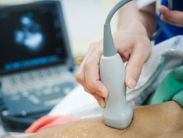
When to use: According to Matthew, ultrasound is commonly used in obstetrics and gynecology to monitor fetal development as well as to evaluate a variety of medical conditions.
“It is ideal for soft tissue evaluation, including monitoring pregnancy, evaluating abdominal organs, detecting gallstones, and examining blood flow in blood vessels. It is also used for guided procedures such as biopsies,” she said.
Who is not suitable: According to Singhealth , ultrasound cannot penetrate bone and cannot show what is inside.
It also cannot pass through air like intestinal gas, so evaluation of the stomach, small intestine, and large intestine may be limited. Deeper parts of the body such as the pancreas and aorta may also be limited, especially if the patient is obese, because body tissue weakens the sound waves as they pass deeper into the body.
4. BREAST IMAGE
What it is: Simply put, it's a specialized X-ray of the breast. “It can identify abnormalities in the breast before symptoms appear, significantly improving treatment outcomes,” says Matthew.
The scan itself is actually very quick, usually taking just a few seconds, according to Dr. Lee. “However, it takes time to position the breast to get the best image, and this can take an additional 5 to 10 minutes, depending on how many additional images are needed.”
“In addition, because compression is required to the breast tissue, the patient may feel some discomfort,” he emphasized.
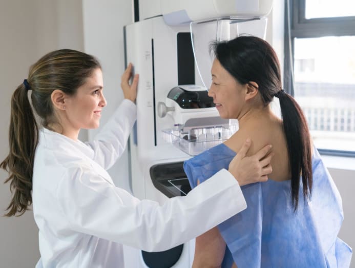
When to use: In addition to routine screening, “mammograms are also used for people who have symptoms like a lump or breast pain to detect any abnormalities,” says Matthew.
Who's not a good candidate: Because of the radiation it uses, the test isn't done in young women until they reach the recommended age for screening, says Dr. Lee.
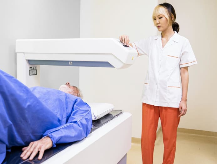
5. BONE DENSITY SCAN
What it is: “This is a very special type of scan that uses X-rays to determine the strength of bones,” says Dr. Lee. The hip or wrist is usually scanned, and it only takes a few minutes to position the body part and perform the scan, he says.
When to use it: “This test is usually done for older patients who are more susceptible to osteoporosis. However, younger patients may be asked to have this test if they are taking certain medications that can affect bone density,” says Dr. Lee.
Who is not suitable: Pregnant women because of the radiation involved. People who have recently had major spinal surgery and those with severe spinal abnormalities such as scoliosis are also excluded because the results may not be accurate.
6. POSIRON EMISSION ATTACK SCAN (PET SCAN)
What it is: This is a highly specialized whole-body scan, according to Dr. Lee. “A special radioactive dye is injected and the patient lies in a scanner, which detects the dye as it is absorbed by different organs.”
He stressed that the process takes about two to three hours because the dye needs time to be absorbed by different organs before the scan is performed.
When it's used: The primary purpose of this scan is to detect cancer and the spread of cancer, but it can also be used to find sources of infection, says Dr. Lee.
Who is not suitable: Dr. Lee says this scan is generally not recommended for children and pregnant patients because of the radiation involved.
Source: CNA/bk
-------------------------------------------------------------------------------------------
👉 Contact SunCare for medical support and advice as well as professional private jet transportation services 🇸🇬 SUNCARE PTE. LTD SINGAPORE
🏠 Add: 10 Anson Road, #10-11 International Plaza, Singapore 079903
☎️ Hotline: +65 96727717 (Dr. Lien Minh - Director) Zalo, Viber
📨 Email: suncarehealth@gmail.com